|

by Mark Sircus
Director
30 May 2011
from
IMVA Website

Homegrown Food
It is in a list of medicinals that
prevent and treat
cancer that we find helpful substances that treat
and strengthen us against radiation contamination.
“In the years leading up to
Chernobyl, some dairy farmers in Austria were using
remineralization as a part of their operations. They added rock
dust to liquid manure as well as combining it with compost,
thereby removing odors and greatly increasing soil biota.
As a result, cows had twice the
normal lifespan and produced much more milk. Amazingly enough,
after Chernobyl, the cheeses that were remineralized (as well as
biodynamic cheeses) measured no radioactivity whatsoever.
Austrians would stand in long lines
in order to buy these safe, remineralized products,”
writes
Joanna Campe.
Iodine is obviously not the only
substance that we should run to in the face of increasing radiation
threats.
Magnesium is a vital mineral whose lack
leaves us open to not only radioactive damages but also those from
heavy metals and thousands of chemicals, which we are commonly
exposed to. Mercury and now a long list of radioactive particles are
floating in the environment like invisible clouds that have spread
out everywhere. They are raining down on us, damaging and damning
our future.
We can no longer be passive about
building our defenses against the toxic onslaught.
Without sufficient magnesium, the body
accumulates toxins and acid residues,
degenerates rapidly, and ages prematurely.
Just about everyone who is writing
protocols for radiation toxicity is forgetting about the importance
of magnesium salts.
Worse still are governments and the entire
institution of medicine that are purposely ignorant about magnesium,
so they cannot possibly be trusted for valuable health and medical
information that will help us in our time of dire need. The need was
dire before Fukushima but they did not want to admit that; they let
the public get obsessed with CO2 emissions and said nothing about
the mercury.
Now with radioactive nuclides steadily building up in
the background, we are in trouble than any of us care to admit.
Today the situation has gone nuclear and there has never before been
a need so great for detoxification and
chelation.
Magnesium is a crucial factor in the natural self-cleansing and
detoxification responses of the body. Magnesium is also necessary
for effective chelation. It stimulates the sodium potassium pump on
the cell wall and this initiates the cleansing process in part
because the sodium-potassium-ATPase pump regulates intracellular and
extracellular potassium levels.
The healthy cell wall favors intake
of nutrients and elimination of waste products.
The involvement of free radicals in tissue injury induced by
magnesium deficiency [1] causes an accumulation of oxidative
products in heart, liver, kidney, skeletal muscle tissues and in red
blood cells,[2] leaving them more vulnerable to oxidative stress
caused by radiation exposure. Both radiation exposure and heavy
metals produce oxidative stress through the creation of increased
levels of reactive oxygen species (ROS
- oxygen free radicals,
peroxides, and singlet oxygen).
It is known that these increased
levels of intracellular ROS are sufficient to trigger apoptosis
(cell death).
Glutathione is
Magnesium-Dependent
Glutathione protects the cells from oxidative-stress-induced
apoptosis and glutathione levels are magnesium dependent!
“Glutathione is a very important detoxifying agent, enabling the
body to get rid of undesirable toxins and pollutants. It forms a
soluble compound with the toxin that can then be excreted through
the urine or the gut.
The liver and kidneys contain high levels of
glutathione as they have the greatest exposure to toxins.
The lungs
are also rich in glutathione partly for the same reason. Many
cancer-producing chemicals, heavy metals, drug metabolites etc. are
disposed of in this way,” says Dr. Patricia Kongshavn, former
professor, department of medicine at McGill University.
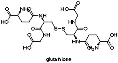
Glutathione is a polypeptide,
(C10H17N3O6S) , of glycine, cysteine, and glutamic acid.
Glutathione synthetase requires glutamyl
cysteine, glycine, ATP, and magnesium ions to form glutathione.[3]
In magnesium deficiency, the ss y-glutamyltranspeptidase is
lowered.[4] There is a direct relationship between cellular
magnesium, GSH/GSSG ratios, and tissue glucose metabolism.[5]
Magnesium deficiency causes glutathione loss and this is unwelcome
as the clouds of radiation are touching down across the northern
hemisphere. Magnesium deficiency causes glutathione loss, which is
not at all healthy because glutathione helps to defend the body
against damage from cigarette smoking, exposure to radiation, cancer
chemotherapy, and toxins such as alcohol and just about everything
else.
According to Dr. Russell Blaylock, low magnesium is associated with
dramatic increases in free radical generation as well as glutathione
depletion and this is vital since glutathione is one of the few
antioxidant molecules known to neutralize mercury.[6]
“For every
molecule of pesticide that your body detoxifies, you throw away or
use up forever a molecule of glutathione, magnesium and more,” says
Dr. Sherry Rogers who goes on to say that, “Your body uses nutrients
to make this glutathione and it uses up energy as well. Every time
we detoxify a chemical, we use up, lose, throw away forever, a
certain amount of nutrients.”
Mineral
Deficiencies
Deficiencies in basic minerals like magnesium and selenium can make
all the difference between health and disease, between being able to
withstand chemical, heavy metal and radiation exposure.
Dr. Rogers
has indicated that there is as much as a 500-fold difference in the
ability of individuals to detoxify the same chemicals and much of
that will be true for radiation as well. A key marker of this
difference is each individual’s magnesium level.
Deficiencies in
magnesium will wreak havoc with our body’s ability to detoxify and chelate heavy radioactive particles and explains much of the
difference between one person withstanding radiation exposures and
another person falling to radiation sickness.
Dr. Leslie Fisher has treated in excess of 35,000 patients where
mineral therapy was prescribed as the sole form of medication.
He
has conducted research within his own clinics and the Department of
Psychiatry, Austin Hospital, Melbourne. Mineral therapy is the
foundation upon which chelation [7] treatments and protocols are
built. Magnesium does protect cells from aluminum, mercury, lead,
cadmium, beryllium and nickel, which explains why re-mineralization
is so essential for heavy metal detoxification and chelation as well
as radiation protection.
Magnesium is essential for the survival of
our cells but takes on further importance now where our bodies are
being bombarded on a daily basis with heavy metals and radiation.
Radiation and
Diabetes
No one is going to convince the public that the increasing radiation
will have a general effect on our health that can be easily traced
back to the source.
Even before we get cancer from radiation we have
a general down-spiraling of body functions because of all the
oxidative stress. In my book, New Paradigms in Diabetes, I write
extensively about the direct relationship between magnesium
deficiency and the onset of diabetes.
Pancreatic beta cells are sensitive to reactive oxygen species (ROS)
[8] attack when they are exposed to oxidative stress,[9] because of
the relatively low expression of antioxidant enzymes such as catalase and glutathione peroxidase.[10]
Diabetes is typically
accompanied by increased production of free radicals and/or impaired
antioxidant defense capabilities, indicating a central contribution
of reactive oxygen species. It is also a fact that ROS is one of the
major factors that induce oxidative modification of DNA and gene
mutation.[11]
The Chernobyl incident was a major disaster of humanity, which has
resulted in a plethora of health problems that are still far from
being fully recognized.
Most studies analyzing the medical
consequences of this catastrophe have so far focused on diseases
such as thyroid cancer, leukemia, immune and autoimmune
pathology[12],[13] even though an increase in the incidence of type
1 diabetes mellitus, a disorder involving the immune system, was
observed within the residential population of Hiroshima among
survivors of the atom bomb detonation.[14]
Studies have also shown
that thymectomy and a sub-lethal dose of gamma radiation induces
type 1 diabetes in rats.[15]
Researchers at the Pediatric Hospital A. Meyer, Florence, Italy
studied this question by assessing the incidence of the disease in
children in Gomel, Belarus in the years subsequent to the Chernobyl
disaster.
The results of the study seem to confirm the hypothesis
that environmental pollution such as that subsequent to the
Chernobyl accident can cause diabetes.[16] Mass screening for diabetes mellitus has been conducted on
64,000-113,000 atomic bomb survivors residing in Hiroshima City
since 1961.
From 1971 to 1992 a 2.7-fold increase in the prevalence
of diabetes mellitus was observed in males and a 3.2-fold increase
in females.[17]
We have a significant and documented
increase in
the incidence of type 1 diabetes in children and
adolescents after Chernobyl in the radioactively
contaminated area of Gomel compared to Minsk.
Heinrich Heine University
When beginning to build a protocol
against the radiation and heavy metal onslaught, we need to stick
with the basics and they are magnesium, iodine, sodium bicarbonate,
vitamin C, selenium, clay, THC (cannabis), as well as a natural
chelator and superfoods.
Properly filtered water also is essential.
There is always more we can do but even
affording these basics is a challenge to many.
Special Note: One
of my readers wrote saying,
“Thank you for the labor of
love you’ve given to the inhabitants of the world. All the
information about magnesium, iodine, baking soda, etc. is
priceless and very much appreciated. I know we can’t thank you
enough for your generosity with the knowledge you’ve accumulated
from all of your research. I know you’ve had your
detractors–ignore them!”
These basic medicines are not
only the mainstay of emergency rooms and intensive care wards but
are the backbone of a my new form of medicine called
Natural
Allopathic Medicine, which makes sense in the age of toxicity that
we all have live in.
Notes
[1] Magnesium deficiency (MgD) has
been associated with production of reactive oxygen species,
cytokines, and eicosanoids, as well as vascular compromise in
vivo. Although MgD-induced inflammatory change occurs during
"chronic" MgD in vivo, acute MgD may also affect the vasculature
and consequently, predispose endothelial cells (EC) to
perturbations associated with chronic MgD. As oxyradical
production is a significant component of chronic MgD, we
examined the effect of acute MgD on EC oxidant production in
vitro. In addition we determined EC; pH, mitochondrial function,
lysosomal integrity and general cellular antioxidant capacity.
Decreasing Mg2+ (< or = 250microM) significantly increased EC
oxidant production relative to control Mg2+ (1000 microM). MgD-induced
oxidant production, occurring within 30 min, was attenuated by
EC treatment with oxyradical scavengers and inhibitors of
eicosanoid biosynthesis. Coincident with increased oxidant
production were reductions in intracellular glutathione (GSH)
and corresponding EC alkalinization. These data suggest that
acute MgD is sufficient for induction of EC oxidant production,
the extent of which may determine, at least in part, the extent
of EC dysfunction/injury associated with chronic MgD. Effect of
acute magnesium deficiency (MgD) on aortic endothelial cell (EC)
oxidant production. Wiles ME, Wagner TL, Weglicki WB.The George
Washington University Medical Center, Division of Experimental
Medicine, Washington, D.C., USA. mwiles@nexstar.com Life Sci.
1997;60(3):221-36.
[2] Martin, Hélène. Richert, Lysiane. Berthelot, Alain Magnesium
Deficiency Induces Apoptosis in Primary Cultures of Rat
Hepatocytes.* Laboratoire de Physiologie, etLaboratoire de
BiologieCellulaire, UFR des Sciences Médicales et
Pharmaceutiques, Besançon, France. 2003 The American Society for
Nutritional Sciences J. Nutr. 133:2505-2511, August 2003.
[3] Virginia Minnich, M. B. Smith, M. J. Brauner, and Philip W.
Majerus.Glutathione biosynthesis in human erythrocytes.
Department of Internal Medicine, Washington University School of
Medicine, J Clin Invest. 1971 March; 50(3): 507–513. Abstract:
The two enzymes required for de novo glutathione synthesis,
glutamyl cysteine synthetase and glutathione synthetase, have
been demonstrated in hemolysates of human erythrocytes. Glutamyl
cysteine synthetase requires glutamic acid, cysteine, adenosine
triphosphate (ATP), and magnesium ions to form ?-glutamyl
cysteine. The activity of this enzyme in hemolysates from 25
normal subjects was 0.43±0.04 ?moleglutamyl cysteine formed per
g hemoglobin per min. Glutathione synthetase requires ?-glutamyl
cysteine, glycine, ATP, and magnesium ions to form glutathione.
The activity of this enzyme in hemolysates from 25 normal
subjects was 0.19±0.03 ?mole glutathione formed per g hemoglobin
per min. Glutathione synthetase also catalyzes an exchange
reaction between glycine and glutathione, but this reaction is
not significant under the conditions used for assay of
hemolysates. The capacity for erythrocytes to synthesize
glutathione exceeds the rate of glutathione turnover by
150-fold, indicating that there is considerable reserve capacity
for glutathione synthesis. A patient with erythrocyte
glutathione synthetase deficiency has been described. The
inability of patients’ extracts to synthesize glutathione is
corrected by the addition of pure glutathione synthetase,
indicating that there is no inhibitor in the patients’
erythrocytes.
[4] Braverman, E.R. (with Pfeiffer, C.C.)(1987). The healing
nutrients within: Facts, findings and new research on amino
acids. New Canaan: Keats Publishing.
[5] Barbagallo, M. et al. Effects of glutathione on red blood
cell intracellular magnesium: relation to glucose metabolism.
Hypertension. 1999 Jul;34(1):76-82. Institute of Internal
Medicine and Geriatrics, University of Palermo, Italy.mabar@unipa.it
[6] http://www.dorway.org/blayautism.txt
[7] Chelation is a recognized treatment for heavy metal
poisoning (such as lead and mercury)
[8] ROS (Reactive Oxygen Species) are natural byproducts of
oxygen metabolism in the body. Free radicals and other
byproducts are formed as a result of this metabolism, and at
lower levels can be very beneficial, but when too many of these
byproducts are formed the situation of oxidative stress occurs.
Reactive oxygen species (ROS) include oxygen ions, free radicals
and peroxides both inorganic and organic. They are generally
very small molecules and are highly reactive due to the presence
of unpaired valence shell electrons. Oxidative stress is a
medical term for damage to animal or plant cells (and thereby
the organs and tissues composed of those cells) caused by
excesses of these reactive oxygen species, which include (but
are not limited to) superoxide, singlet oxygen, peroxynitrite or
hydrogen peroxide. Superoxide is produced deleteriously by
1-electron transfers in the mitochondrial electron transfer
chain. It is defined as an imbalance between pro-oxidants and
anti-oxidants, with the former prevailing. The causes of these
excesses are many, and include environmental influences of every
type. Enzyme activities are sometimes affected negatively,
leading to greater production of excess ROS, and heavy metals
such as chromium, vanadium, and others are said to be involved,
now this new evidence that methylmercury definitely plays a
significant role in the pancreas. Cells are normally able to
defend themselves against ROS damage through the use of enzymes
such as superoxide dismutases and catalases. Small molecule
antioxidants such as Ascorbic acid (vitamin-C), uric acid, and
glutathione also play important roles as cellular antioxidants.
Similarly, Polyphenol antioxidants assist in preventing ROS
damage by scavenging free radicals. Studies are conflicting on
some antioxidants such as Vit. E. The resulting inflammatory
processes are believed to be the result of these ROS excesses
and include cardiovascular disease, ALS, neurodegenerative
diseases, and many others.
[9] Kajimoto, Y., and Kaneto, H. (2004) Role of oxidative stress
in pancreatic beta-cell dysfunction. Ann. N. Y. Acad. Sci. 1011,
168-176.
[10] Tiedge, M., Lortz, S., Drinkgern, J., and Lenzen, S. (1997)
Relation between antioxidant enzyme gene expression and
antioxidative defense status of insulin-producing cells.
Diabetes 46, 1733-1742.
[11] Inoue, M., Sato, E. F., Nishikawa, M., Hiramoto, K.,
Kashiwagi, A., and Utsumi, K. (2004) Free radical theory of
apoptosis and metamorphosis.Redox Rep. 9, 237-247.
[12] Kuzmenok O, Potapnev M, Potapova S et al. (2003) Late
effects of the Chernobyl radiation accident on T cell-mediated
immunity in cleanup workers. Radiat Res 159: 109–116.
[13] Lomat L, Galburt G, Quastel MR, Polyakov S, Okeanov A,
Rozin S (1997) Incidence of childhood disease in Belarus
associated with the Chernobyl accident. Environ Health Perspect
[Suppl 105] 6:1529–1532.
[14] Ito C (1994) Trends in the prevalence of diabetes mellitus
among Hiroshima atomic bombsurvivors. Diabetes Res ClinPract [Suppl]:S29–S35.
[15] Ramanathan S, Bihoreau MT, Paterson AD, Marandi L, Gauguier
D, Poussier P (2002) Thymectomy and radiationinduced type 1
diabetes in nonlymphopenic BB rats. Diabetes 51:2975–2981.
[16] J PediatrEndocrinolMetab. 2002 Jan;15(1):53-7. Incidence of
childhood type 1 diabetes mellitus in Gomel, Belarus.Martinucci
ME, Curradi G, Fasulo A, Medici A, Toni S, Osovik G, Lapistkaya
E, Sherbitskaya E. Regional Centre for Juvenile Diabetes,
Paediatric Hospital A. Meyer, Florence, Italy.
[17] Trends in the prevalence of diabetes mellitus among
Hiroshima atomic bomb survivors. Diabetes Res ClinPract. 1994
Oct;24 Suppl:S29-35. Hiroshima Atomic Bomb Casualty Council,
Health Management Center, Japan.
|