|
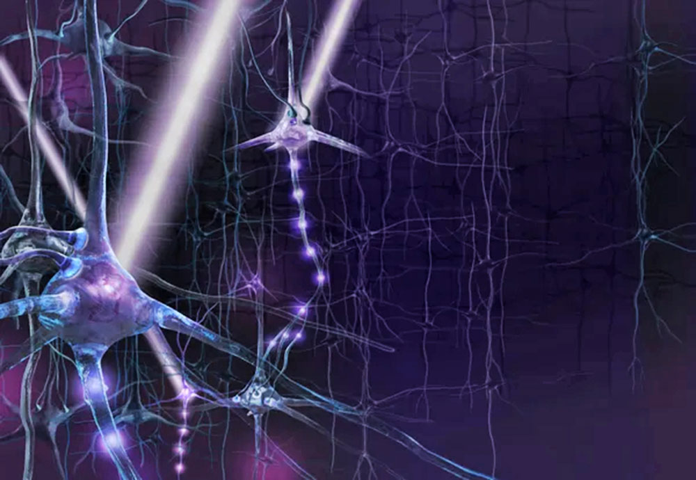
Getty Images the author offers a longer version of his December 2010 Scientific American article on how researchers can probe how the nervous system works in unprecedented detail, using a technique called optogenetics
Clearly, we need new answers in psychiatry.
But
as philosopher of science
Karl Popper might have said, before
we can find the answers, we need the power to ask new questions. In
other words, we need new technology.
Because of that complexity, neuroscientists lack a deep grasp of what the brain is really doing - of how specific activity patterns within specific brain cells ultimately give rise to thoughts, feelings and memories. By extension, we also do not know how the brain's physical failures produce distinct psychiatric disorders such as depression or schizophrenia.
The ruling paradigm of psychiatric disorders - casting them in terms of chemical imbalances and altered levels of neurotransmitters - does not do justice to the brain's high-speed electrical neural circuitry.
And psychiatric treatments have historically been
largely serendipitous: helpful for many but rarely illuminating, and
suffering from the same challenges as basic neuroscience.
Electrical stimuli cannot meet this challenge because electrodes are too crude a tool: they stimulate all the circuitry at their insertion site without distinguishing between different cell types, and their signals cannot turn off neurons with precision.
Drugs are not specific enough either, and they are much slower than the natural operating speed of the brain.
Crick later speculated in lectures that light
might have the properties to serve as a control tool because it
could be delivered in precisely timed pulses, but at the time no one
had a strategy to make specific cells responsive to light.
At least 40 years ago biologists knew that some microorganisms produce proteins that directly regulate the flow of electric charge across cell membranes in response to visible light. These proteins, which are produced by a characteristic set of "opsin" genes, help to extract energy and information from the light in the microbes' environments.
In 1971 Walther Stoeckenius and Dieter Oesterhelt, both then at the University of California, San Francisco, discovered that one of these proteins, bacteriorhodopsin, acts as a single-component ion pump that can be briefly activated by photons of green light - a remarkable all-in-one molecular machine.
Later identification of other members of this
family of proteins - the halorhodopsins in 1977 and the
channelrhodopsins in 2002 - continued this original theme from 1971
of single-gene, all-in-one control.
Yet it took more than 30 years, until the summer
of 2005, for these fields to come together in a new technology (optogenetics)
based on microbial opsin genes.
It includes the discovery and insertion into
cells of genes that confer light responsiveness; it also includes
the associated technologies for delivering light deep into organisms
as complex as freely moving mammals, for targeting light-sensitivity
to cells of interest, and for assessing specific readouts, or
effects, of this optical control.
The significance of any event in a cell has full meaning only in the context of the other events occurring around it in the rest of the tissue, the whole organism or even the larger environment.
Even a shift of a few milliseconds in the timing of a neuron's firing, for example, can sometimes completely reverse the effect of its signal on the rest of the nervous system.
And millisecond-scale timing precision within
behaving mammals has been essential for key insights into both
normal brain function and into clinical problems such as
parkinsonism.
Even a single psychiatric disease, major depression, is the leading cause of disability worldwide in women aged 15 to 44. But much stigma remains (which may relate to why hearing about this epidemiology is so surprising to many people).
Why the stigma?
A major reason is our collective lack of understanding.
Just as a cancer diagnosis once carried more stigma than it does now (perhaps because of confusion over what cancer really "is," over concerns for contagion or even over blame for the cancer on personality features of the patient), so too does lack of insight into psychiatric disease contribute to stigmatization, further slowing progress in this enormous problem for global human health.
This lack of insight, sadly, is universal:
throughout the global community, from members of the general public
to the most influential and advanced psychiatrists, we don't know
what psychiatric disease "is" at a fundamental level.
I come face-to-face with this challenge continually. In addition to running a research laboratory in a bioengineering department, I am also a practicing psychiatrist, and I treat patients regularly using combinations of medication, therapy, and electrical or magnetic brain stimulation.
After my undergraduate years at Harvard University, I had obtained my MD and PhD degrees at Stanford University, focusing on synaptic electrophysiology and optical studies of mammalian neural circuitry.
I then completed my psychiatry residency and postdoctoral fellowships at Stanford, where I developed as a physician and developed skills in the study of animal behavior.
Although as a physician I employ modern tools (such as transcranial magnetic stimulation), these tools are still not good enough and, most important, do not provide deep insight into the diseases, only highlighting (as do the patients) our limitations. I remember a brilliant young college student suffering from psychotic depression and terrified by the incomprehensible voices and uncontrollable bizarre ideas in his mind.
I remember a retired woman so severely depressed that she was unable to smile, barely able to eat and unresponsive to her grandchildren.
My inability to explain these changes in a
scientific way and the unfortunately failed responses to treatments
these patients experienced have never left my mind.
Being asked to reflect on our optogenetics
work here also provides an opportunity to consider broader
implications of the scientific process.
Researchers have long employed a light-based method called CALI to destroy, and thus inhibit, selected proteins; lasers have also been used to destroy specific cells, for example, in the worm Caenorhabditis elegans.
Conversely, Richard L. Fork of Bell Laboratories (in the 1970s) and Rafael Yuste of Columbia University (in 2002) reported ways to stimulate neurons with lasers that partially disrupted cell membranes.
More recently, the laboratories of Gero Miesenböck, then at Memorial Sloan–Kettering Cancer Center, and of Ehud Isacoff, Richard H. Kramer and Dirk Trauner, then all at the University of California, Berkeley, employed multicomponent systems for modulating targeted cells with light.
They introduced, for example, both a protein that
regulates neurons and a chemical that would spur the protein into
action when triggered by ultraviolet light.
A fundamental strategic shift to a single-component strategy was necessary.
As it turned out, this single-component strategy
was not able to build on any of the parts or methods from earlier
approaches, but instead employed the remarkable all-in-one
light-activated proteins from microbes: proteins now called
bacteriorhodopsins, halorhodopsins and channelrhodopsins.
While reviewing them, Peter Hegemann, then at the University of Regensburg in Germany, who had predicted that Chlamydomonas would have a light-activated ion channel, noticed two long sequences similar to those for bacteriorhodopsin. He obtained copies of them from Kazusa and asked Georg Nagel (then a principal investigator in Frankfurt) to test if they indeed coded for ion channels.
In 2002 Hegemann and Nagel described their finding that one of these sequences encoded a single-protein membrane channel responsive to blue light: when hit by blue photons, it regulated the flow of positively charged ions.
The protein was consequently dubbed channelrhodopsin-1, or ChR1.
The following year Nagel and Hegemann (along with their colleagues, including Ernst Bamberg in Frankfurt) explored the other sequence and named the encoded protein "channelrhodopsin-2," or ChR2.
Almost simultaneously, John L. Spudich in Houston provided evidence that those genes were important to the light-dependent responses of Chlamydomonas.
But these channelrhodopsins - a third type of
single-component light-activated ion-conductance protein - did not
immediately translate into an advance in neuroscience any more than
the discoveries of bacteriorhodopsins and halorhodopsins in previous
decades had. Several years passed uneventfully after 2002, as they
had since 1971.
Animal cells were unlikely to manufacture these microbial membrane proteins efficiently or safely, and the proteins were virtually certain to be too slow and weak to be effective.
Furthermore, to function, the proteins would
require an additional cofactor - a vitamin A–related compound called
all-trans retinal to absorb the photons. The risk of wasting time
and money was far too great.
As a principal investigator at Stanford beginning in 2004, I formed a team that included the extraordinarily talented graduate students Edward Boyden and Feng Zhang (both now assistant professors at the Massachusetts Institute of Technology) to address this challenge.
I introduced channelrhodopsin-2 into mammalian neurons in culture by the well-established techniques of transfection - that is, by splicing the gene for ChR2 and a specific kind of on switch, or promoter, into a vector (like a benign virus) that ferried the added genetic material into the cells.
Promoters can ensure that only selected kinds of
neurons (such as those able to secrete the neurotransmitter
glutamate) will express, or make, the encoded proteins.
In August 2005 my team published the first report that by introducing a microbial opsin gene, we could make neurons precisely responsive to light. Channelrhodopsins (and, eventually as we found, the bacteriorhodopsin from 1971 and the halorhodopsins, too) all proved able to turn neurons on or off, efficiently and safely in response to light.
They worked in part because mammalian tissues contain naturally robust quantities of all-trans retinal - the one chemical cofactor essential for photons to activate microbial opsins - so nothing beyond an opsin gene needs to be added to targeted neurons.
Microbial opsin genes provided the long-sought single-component strategy.
Improving on Nature
Investigators are adding new opsins to their tool kits by scouring the natural world for novel
ones; they are also applying molecular engineering to tweak the
known opsins to make them even more useful for diverse experiments
in a wider range of organisms.
Using VChR1 and the other channelrhodopsins together, we can simultaneously control mixed populations of cells, with yellow light exerting one type of control over some of them and blue light sending a different command to others. And we now have found that the most potent channelrhodopsin of all is actually a hybrid of VChR1 and ChR1 (with no contribution from ChR2 at all).
Our other modified opsins (created with Ofer Yizhar, Lief Fenno, Lisa Gunaydin and Hegemann and his students) now include "fast" and "slow" channelrhodopsin mutants that offer exquisite control over the timing and duration of action potentials: the former can drive action potentials more than 200 times per second, whereas the latter can push cells into or out of stable excitable states with single pulses of light.
Our newest opsins can also now respond to deep red light that borders on the infrared, which stays more sharply focused, penetrates tissues more easily and is very well tolerated by subjects.
Many groups are now also pushing opsin
engineering forward, including those of Hiromu Yawo in Japan,
Ernst Bamberg in Frankfurt and Roger Tsien in San
Diego.
For example, pieces of "trafficking" DNA can be
bundled with the opsin genes to act as "zip codes" to ensure the
genes are transported to the correct compartments within mammalian
cells and translated properly into functional proteins. This
generalizable approach has served to unlock the broad ecological
repertoire of microbial opsin genes.
These proteins sense extracellular signaling chemicals, such as epinephrine, and respond by changing the levels of intracellular biochemical signals, such as calcium ions, and thus the activity of the cells.
By adding the light-sensing domain from a rhodopsin molecule to G-protein coupled receptors, early in 2009 Raag Airan and others in my laboratory published a set of receptors called optoXRs that respond rapidly to green light.
When viruses are used to insert the
single-component optoXR genes into the brains of lab rodents, the
first cell type–specific fast optical control over defined
biochemical pathways was enabled, working even in freely moving
mammals. Optical control over defined biochemical events is now also
being explored in many laboratories, and opens the door to
optogenetics in essentially every cell and tissue in biology.
And to enable simultaneous readouts of the dynamic electrical signals elicited by optogenetic control, we also have published millisecond-scale instruments that are integrated hybrids of fiber optics and electrodes ("optrodes").
A long-sought synergy can emerge between optical stimulation and electrical recording because the two can be set up to not interfere with each other. We can now, for instance, directly observe the changing electrical activity in the neural circuits involved in motor control at the same time that we are optically controlling those circuits with microbial opsins.
The more rich and complex that both our
optogenetic inputs and the electrical-output measures of neural
circuits become, the more powerfully we can infer the computational
and informational roles of neural circuits from how they transform
our signals.
In addition to distributing these diverse engineered opsin genes to more than 700 laboratories worldwide (https://www.optogenetics.org), my students have worked hard over the past few years to develop and deliver optogenetics instruction.
We have found that despite the unusual
combination of technologies required for optogenetics, the
fundamentals can be taught in focused hands-on courses in the
laboratory, which accelerate the benefits of the technology.
Scientists from all over the world come to practice optogenetics and
return to their home institutions, where they serve as local sources
of knowledge and wisdom, resulting in application to diverse
settings and challenges across the globe.
In recent years, neuroscience has made many advances based on the brain-scanning technique called functional magnetic resonance imaging (fMRI). These scans are usually billed as providing detailed maps of neural activity in response to various stimuli.
Yet strictly speaking, fMRI shows only changes in blood-oxygen levels in different areas of the brain; those changes are just a proxy for actual neural activity. Some nagging uncertainty has therefore always surrounded the question of whether these complex fMRI signals can be triggered by increases in local excitatory neural activity.
This past May, however, my laboratory used a combination of optogenetics and fMRI (dubbed ofMRI) to verify that the firing of local excitatory neurons is fully sufficient to trigger the complex signals detected by fMRI scanners.
In addition, ofMRI can map working neural
circuits with an exactness and completeness not previously possible
with electrodes or drugs. Optogenetics is thereby helping to
validate and advance a wealth of scientific literature in
neuroscience and psychiatry.
Optogenetics has also been employed to help determine how dopamine-making neurons may give rise to feelings of reward and pleasure.
In this work with Hsing-chen Tsai, Feng Zhang, Antonello Bonci, Garrett Stuber and Luis de Lecea, we optogenetically drove well-defined dopamine neurons in the mouse in different temporal patterns during free behavior, and found parameters that were sufficient to drive reinforced behavior (for example, in the absence of any other cue or reward, healthy animals simply chose to spend more time in places where they had received particular kinds of optogenetic bursting activity in dopamine neurons).
This work is relevant to hedonic
(pleasure-related) pathologies involved in depression (as in my
depressed patient who could no longer even enjoy seeing her
grandchildren, otherwise one of the most rewarding experiences known
to humankind) and in substance abuse, as well as in healthy reward
processes.
Since the 1990s some Parkinson's patients have received relief via a therapy called deep-brain stimulation, in which an implanted device similar to a pacemaker applies carefully timed, oscillating electric stimuli to certain areas far inside the brain, such as the subthalamic nucleus.
Yet the promise of this technique for Parkinson's (and indeed for a variety of other conditions) is partially limited because electrodes stimulate nearby brain cells unselectively and medical understanding of what stimuli to apply is woefully incomplete.
Recently, however, we have used optogenetics to study animal models of Parkinson's and gained fundamental insight into the nature of the diseased circuitry and the mechanisms of action of therapeutic interventions.
For example, we have found that deep-brain stimulation may be most effective when it targets not cells but rather the connections between cells - affecting the flow of activity between brain regions.
And we have worked with Anatol
Kreitzer of U.C.S.F. who has functionally mapped two pathways in
brain movement circuitry: one that slows movements and one that
speeds them up and can counteract the parkinsonian state.
Using optogenetics, Vikaas Sohal and Feng Zhang in my group (along with Li-Huei Tsai and Chris Moore at M.I.T. and our other collaborators) showed that parvalbumin cells, in cooperation with other cell types, serve to modulate gamma waves.
Those waves in turn enhance the flow of information through cortical circuits. In my patients with schizophrenia I see what clearly appear to be information-processing problems, in which mundane random events are incorrectly viewed as parts of larger themes or patterns (an informational problem perhaps giving rise to paranoia and delusions).
These patients may suffer from some failure of an internal "notification" mechanism that informs us when thoughts that are self-generated (an informational problem perhaps underlying the frightening phenomenon of "hearing voices").
Conversely in my patients with autism spectrum disease, rather than inappropriately broad linkages in information, I see overly restricted information processing: they miss the big picture by focusing too narrowly on just parts of objects, people, conversations and so on.
These failures of information processing may lead
to failures in communication and social behavior, and better
understanding of brain rhythms has provided insights into these
complex diseases.
Optogenetics is thus helping to move psychiatry toward a network-engineering approach, in which the complex functions of the brain (and the behaviors it produces) are interpreted as properties of the neural system that emerge from the electrochemical dynamics of the component cells and circuits. It thus fundamentally changes our understanding of how electrically excitable tissues function in health and disease.
We have come a long way from bacteriorhodopsin,
indeed.
At large and diverse scientific gatherings (such as the Society for Neuroscience annual meeting, with more than 30,000 attendees), I have occasionally heard colleagues overwhelmed by the diversity of the field take the devil's advocate position and suggest that it would be more efficient to focus tens of thousands of scientists on one massive and urgent project at a time - for example, eliminating Alzheimer's disease.
Indeed, a common topic of conversation and policymaking in science considers the extent to which either diverse exploration or massively focused efforts should predominate. Even for small focused efforts, how much should funding sources and scientific organizations set the scientific agenda and guide investigators?
There is no question that both directed and
exploratory approaches have value, and as a practicing psychiatrist
I fully appreciate the urgency for directed efforts. As they did for
me, clinical need can and should drive and inspire basic science and
engineering.
The story of optogenetics makes a strong argument. Even envisaging the concept of optical control of targeted neurons, as Crick did, did not predict the need for decades of pure basic research on microbial membranes.
The bottom line is that there is no possible way one could have predicted the impact of archaeal or algal biology on our understanding of Parkinson's disease and its treatment.
It is still too early in the exhilarating history
of humanity's march toward understanding of the natural world.
Purely directed funding will adversely affect most seriously the fields most distant from human health (such as archaeal biology), but will even stultify fields clearly relevant to human health and disease, such as neuroscience.
In a supreme irony,
even if 30,000 scientists could be directed toward a common goal,
the very act of focusing on that goal could ensure that the goal
would not be reached.
Consider the importance of basic science - which could be so vulnerable to criticism when misrepresented - in the case of optogenetics, which could be cast in an unflattering light as money spent studying genes from pond scum.
In classic microbial opsin work, we find meaning for the modern world - not just for science, but also for medicine and psychiatry - that makes a strong and clear statement for environmental protection, for preservation of biodiversity and for the pure quest for understanding.
And the journey of optogenetics shows that hidden within the ground we have already traveled over or passed by, there may reside the essential tools, shouldered aside by modernity, that will allow us to map our way forward.
Sometimes these neglected or archaic tools are
those that are most needed - the old, the rare, the small and the
weak.
Kennedy concluded,
I would add to that magnificent sentiment only that the story of optogenetics recounted above sends a strong, clear message. In our quest to move the enterprise of science forward we should never forge
|