|

by Carissa Wong
May 09, 2024
from
Nature Website
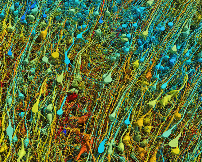
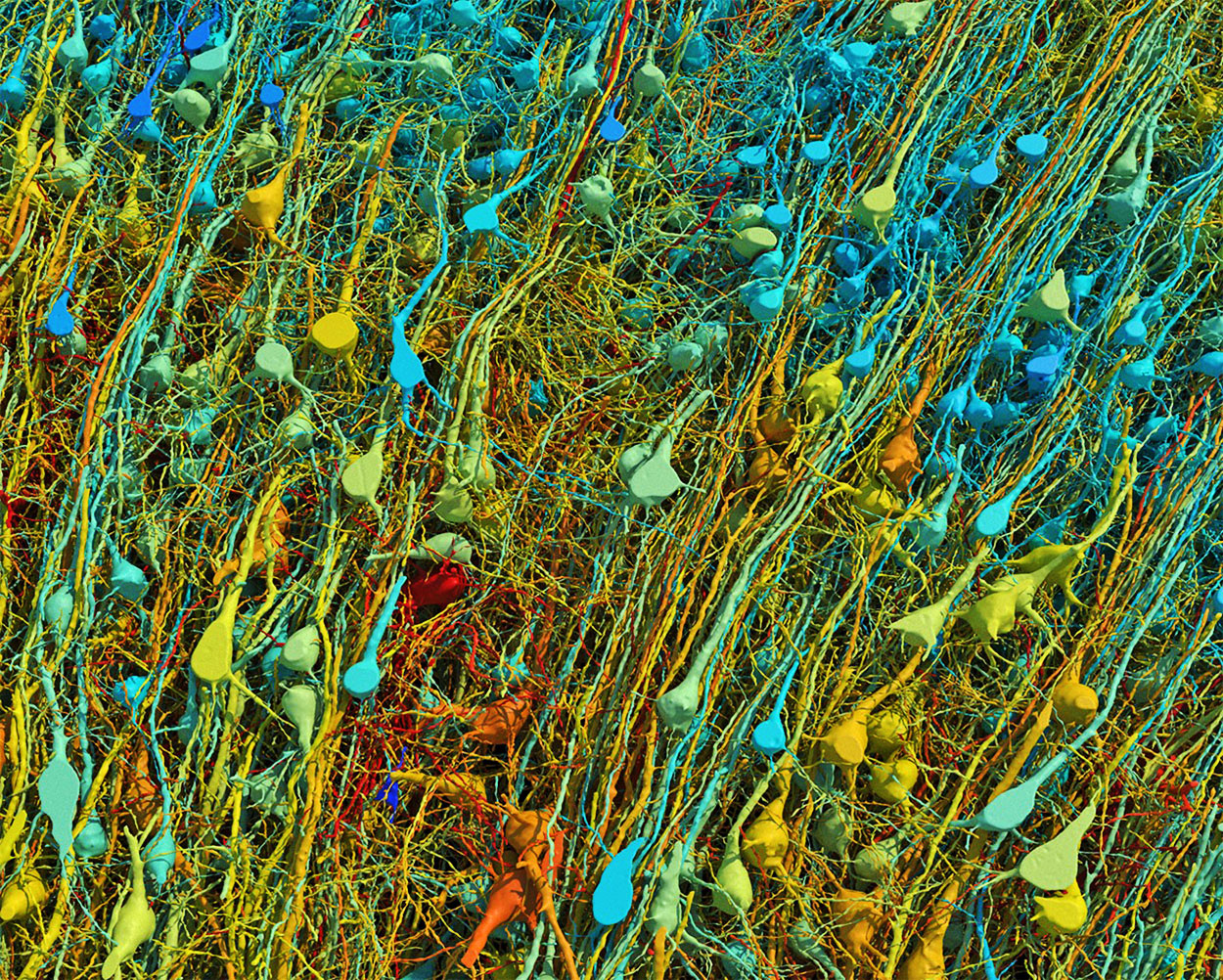
Rendering based on
electron-microscope data,
showing the
positions of neurons in a fragment of the brain cortex.
Neurons are
colored according to size.
Credit: Google
Research & Lichtman Lab (Harvard University).
Renderings by
D. Berger (Harvard University)
Google scientists have modeled a fragment
of the
human brain at nanoscale resolution,
revealing
cells with previously undiscovered features...
Researchers have mapped a tiny piece of the human brain in
astonishing detail.
The resulting cell atlas, which was described
today in Science 1 and is available online,
reveals new patterns of connections between brain cells called
neurons, as well as cells that wrap around themselves to form knots,
and pairs of neurons that are almost mirror images of each other.
The 3D map covers a volume of about one cubic millimeter,
one-millionth of a whole brain, and contains roughly 57,000 cells
and 150 million synapses - the connections between neurons.
It incorporates a colossal 1.4 petabytes of data.
"It's a little bit humbling," says Viren
Jain, a neuroscientist at Google in Mountain View,
California, and a co-author of the paper.
"How are we ever going to really come to
terms with all this complexity?"
Slivers of brain
The brain fragment was taken from a 45-year-old woman when she
underwent surgery to treat her epilepsy.
It came from the cortex, a part of the brain
involved in learning, problem-solving and processing sensory
signals. The sample was immersed in preservatives and stained with
heavy metals to make the cells easier to see.
Neuroscientist Jeff Lichtman at Harvard
University in Cambridge, Massachusetts, and his colleagues then cut
the sample into around 5,000 slices - each just 34 nanometres thick
- that could be imaged using electron microscopes.
Jain's team then built artificial-intelligence models that were able
to stitch the microscope images together to reconstruct the whole
sample in 3D.
"I remember this moment, going into the map
and looking at one individual synapse from this woman's brain,
and then zooming out into these other millions of pixels," says
Jain.
"It felt sort of spiritual."
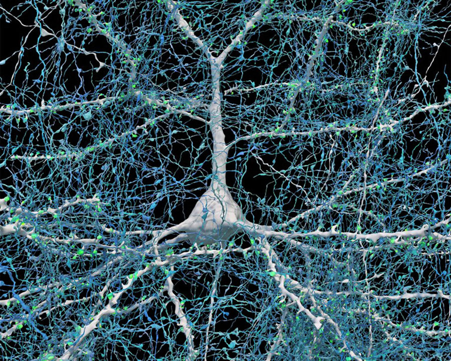
Rendering of a neuron with a round base and many
branches, on a black background.

A single neuron
(white)
shown with 5,600 of
the axons (blue) that connect to it.
The synapses that
make these connections
are shown in green.
Credit: Google
Research & Lichtman Lab (Harvard University).
Renderings by D.
Berger (Harvard University)
When examining the model in detail, the researchers discovered
unconventional neurons, including some that made up to 50
connections with each other.
"In general, you would find a couple of
connections at most between two neurons," says Jain.
Elsewhere, the model showed neurons with tendrils
that formed knots around themselves.
"Nobody had seen anything like this before,"
Jain adds.
The team also found pairs of neurons that were
near-perfect mirror images of each other.
"We found two groups that would send their
dendrites in two different directions, and sometimes there was a
kind of mirror symmetry," Jain says.
It is unclear what role these features have in
the brain.
Proofreaders needed
The map is so large that most of it has yet to be manually checked,
and it could still contain errors created by the process of
stitching so many images together.
"Hundreds of cells have been 'proofread', but
that's obviously a few per cent of the 50,000 cells in there,"
says Jain.
He hopes that others will help to proofread parts
of the map they are interested in.
The team plans to produce similar maps of brain
samples from other people - but a map of the entire brain is
unlikely in the next few decades, he says.
"This paper is really the tour de force
creation of a human cortex data set," says Hongkui Zeng,
director of the Allen Institute for Brain Science in Seattle.
The vast amount of data that has been made freely
accessible will,
"allow the community to look deeper into the
micro-circuitry in the human cortex", she adds.
Gaining a deeper understanding of how the cortex
works could offer clues about how to treat some psychiatric and
neurodegenerative diseases.
"This map provides unprecedented details that
can unveil new rules of neural connections and help to decipher
the inner working of the human brain," says Yongsoo Kim,
a neuroscientist at Pennsylvania State University in Hershey.
References
-
Shapson-Coe, A. et al. Science 384,
eadk4858 (2024)
|