|
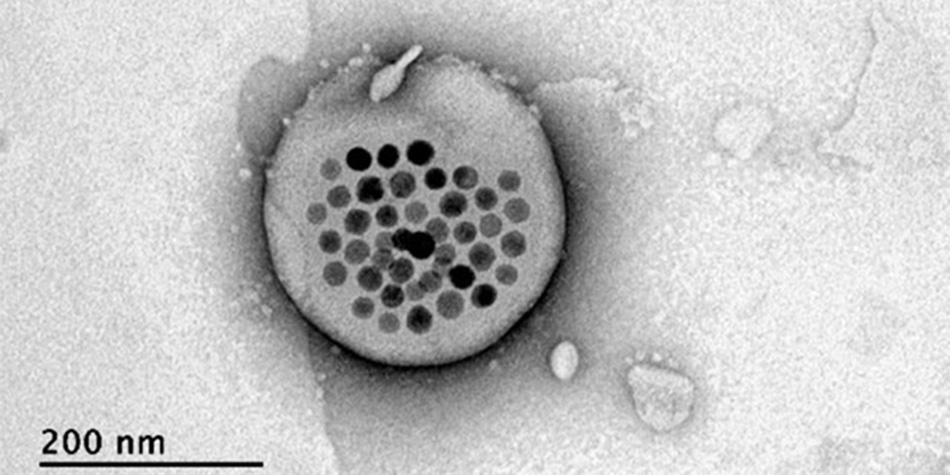
showing iron oxide particles inside a liposome "bubble". (MIT)
A team of MIT scientists
has constructed a type of heat-sensitive, magnetic nanoparticle that
can deliver chemical stimulants deep into brain tissues and release
them on demand, providing a new means to remotely modulate the
behaviors of test subjects.
Due to their biocompatibility, ability to
entrap a variety of small and large molecules, and versatility to
adopt a wide range of physicochemical and biological properties, liposomes are a popular carrier in biomedical science, capable of
delivering anything from plasmid DNA for gene editing, to cytotoxic
chemo-agents in cancer therapy.
They are not only a good contrast agent in Magnetic Resonance Imaging (MRI) scans, but also a perfect vehicle to induce magnetic hyperthermia - a technique extensively used in oncology treatment.
In a typical procedure, a colloid preparation made of nanoscale iron oxide compounds is injected directly to the vein that feed a tumor.
The colloidal particles get heated up upon
exposure to an alternating high-frequency magnetic field, which
enables them to "bake" and eventually kill off cancerous tissues
within the tumor.
DBS, which
involves placing stimulating electrodes deep inside a subject's
brain, is effective in treating neurodegenerative disorders such as
Parkinson’s disease and essential tremor.
They utilized a so-called
magnetogenetic
approach - essentially deploying blood-brain barrier (BBB)-crossing MNPs to the targeted brain region and using the thermal energy
generated by magnetic hyperthermia to release chemical stimulants
encapsulated inside these lipid bubbles.
About 20 seconds later,
when the liposomal particles reached a temperature of 42º celsius (107.6° F), the entrapped drug molecules were
seen escaping from the thermally sensitive MNP.
Check out this video below from Nanoprobes Inc.:
|