|

by Enrico Trigoso
September 21, 2022
from
TheEpochTimes Website
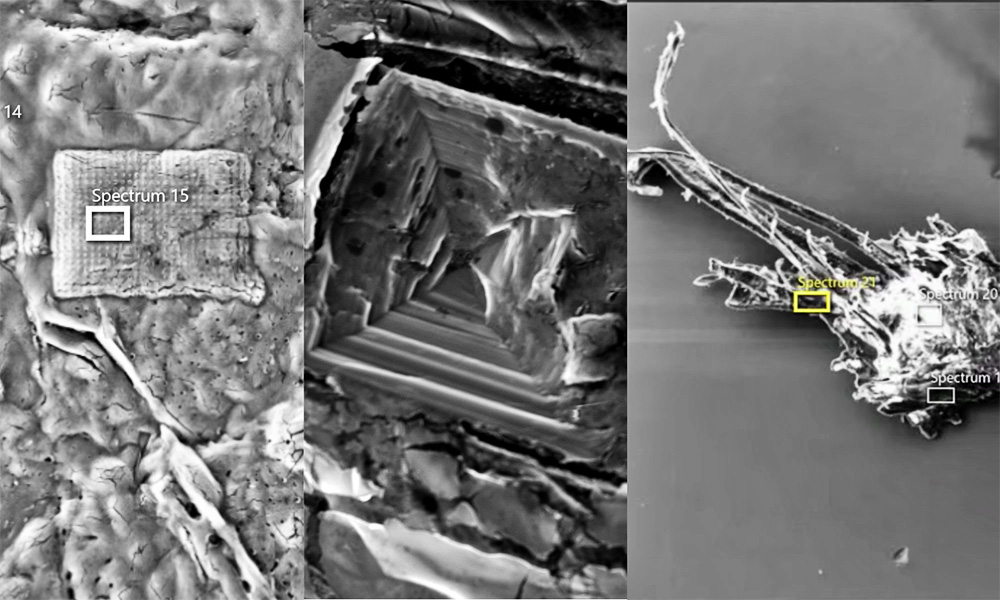
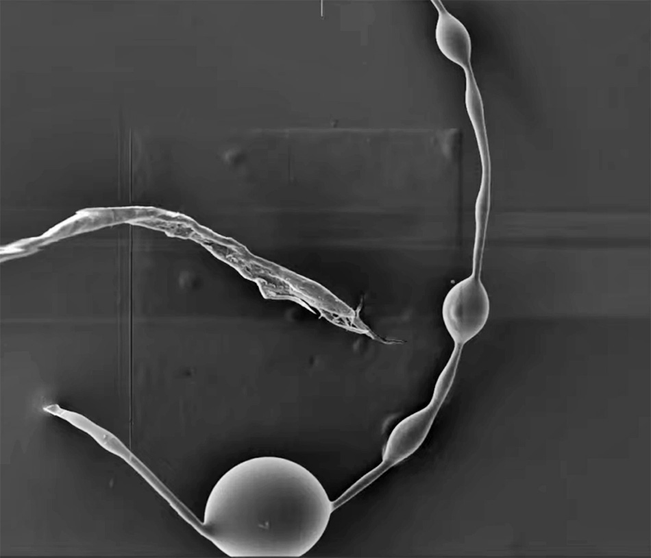
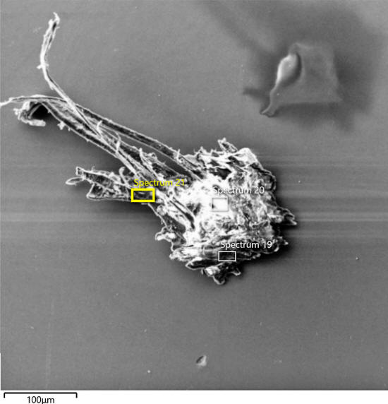
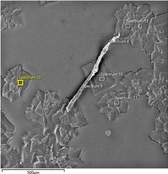
SEM (Scanning Electron Microscope) image
from vaccine vials.
(Courtesy of Dr. Daniel Nagase)
Dr.
Daniel Nagase has been using a scanning electron
microscope to analyze both
Pfizer and Moderna mRNA vaccines
that were exposed to room temperature for weeks or months, and has
found odd objects that according to several doctors who talked to
us, should not be in the vials - even after degradation...
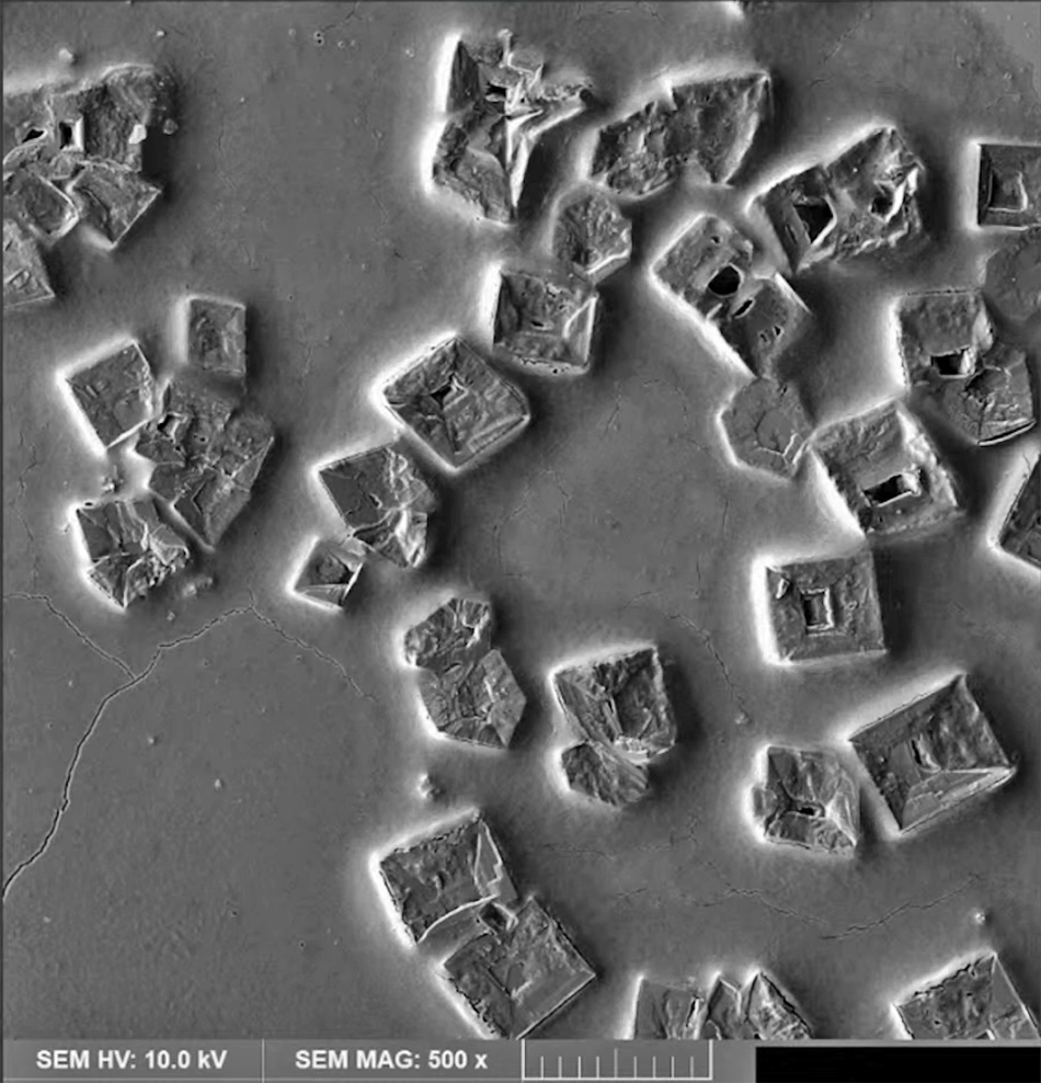
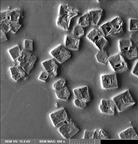
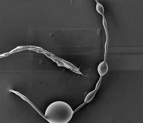
Nagase took photos of crystals, spheres, fibers, and most
strikingly:
"rectangles and
inverted pyramids."
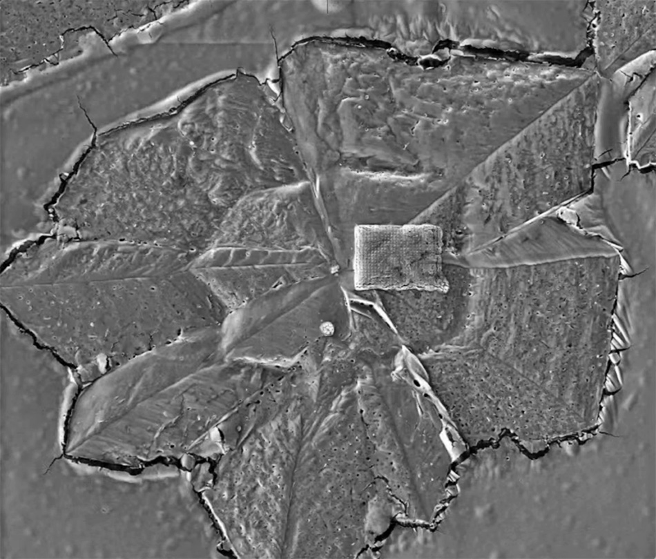
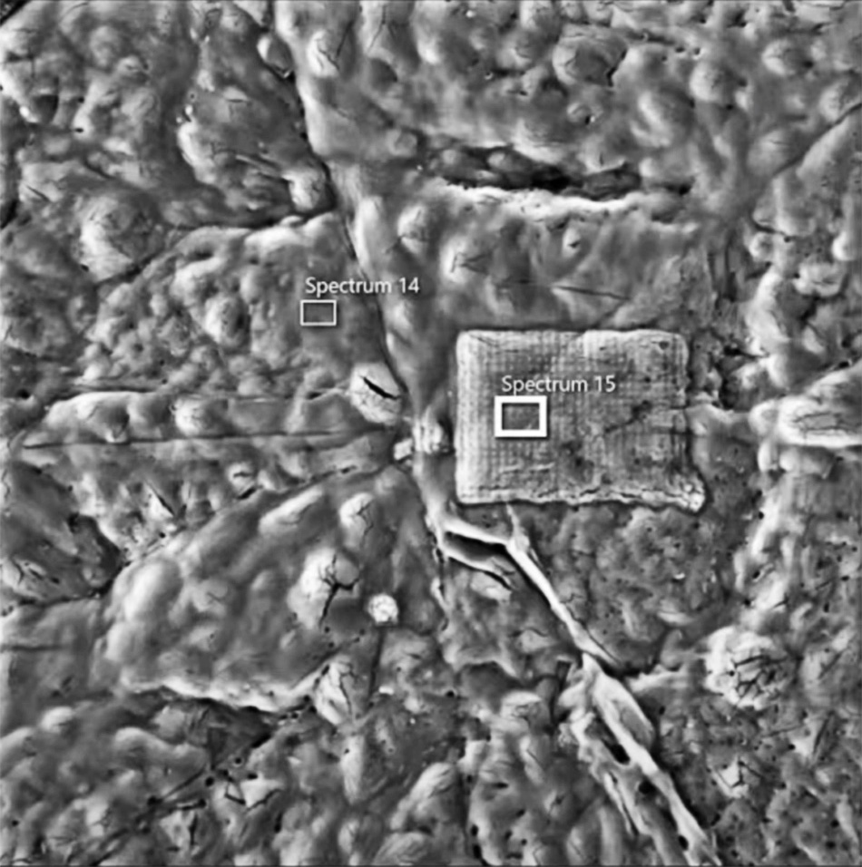
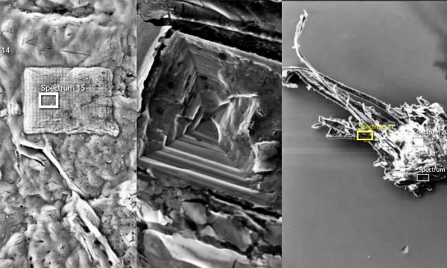
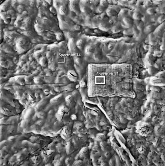
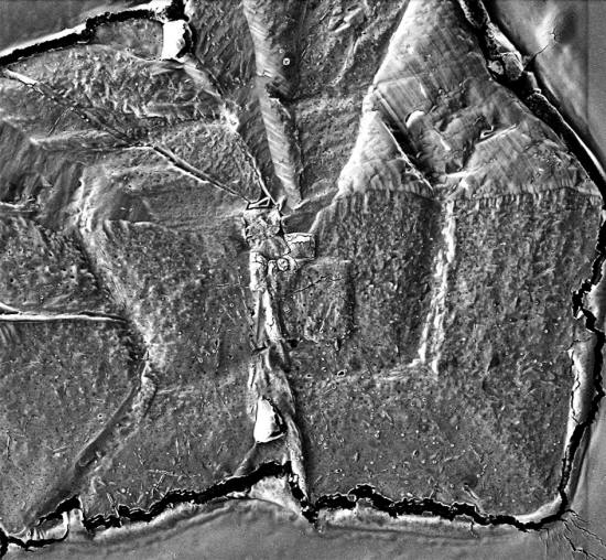
One of the images shows
a,
"hexagonal
crystalline structure, on top of which there is a 4-sided
rectangular structure with regularly spaced dots in the form of
a grid."
"4-sided structures on top of 6-sided structures do not occur
naturally," Nagase told us, "neither do grid markings."
A molecular
biologist/virologist who reviewed Nagase's findings told us under
the condition of anonymity that the results of the scanning electron
microscopy "revealed unexpected content."
"Most notable is a
distinct rectangular shape consisting primarily of carbon and
oxygen," the scientist told us.

SEM (Scanning Electron Microscope)
image from Pfizer vial, March batch.
(Courtesy of Dr. Daniel Nagase)
"This shape is
inconsistent with known morphological characteristics of vaccine
components and biological matter.
Further investigation
of vaccine composition is pertinent and independent verification
in controlled conditions is urgently needed."
No Nitrogen,
No Phosphorous in First Batch
Nagase's main concern is that they are composed of "just carbon and
oxygen."
He maintains that biologics such as RNA would show signs of nitrogen
and phosphorous, but his first batch (from March) of microscopic
scans of Pfizer and Moderna vials did
not show either of these elements on his machine.
The August batch did detect phosphorous,
"indicating there is
either DNA or RNA in the sample."
The scanning electron
microscope he uses is able to - and has detected - nitrogen in other
different scans which he showed to us.
He did not want to reveal the brand of the apparatus because,
"that might identify
the machine."
Nagase is an ER Doctor
from Canada who has been practicing since 2008.
He studied Physiology and
Cell Biology at McGill University and also went to Dalhousie
University Medical School.
The
type of microscope Nagase is using
is able to shoot an electron beam through electromagnetic fields and
lenses and concentrates the beam toward the object that is being
analyzed, then the beam bounces outward with electrons and X-rays,
and shows what chemical elements the object was composed of.
He has done two series of analyses on the mRNA vaccines.
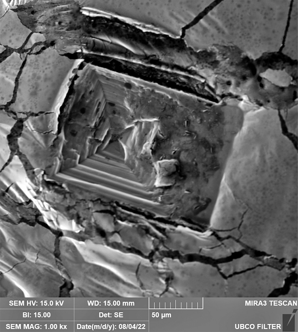
The last one was done
around four months ago, and the new one was done in August. He has
sent us a new image (the inverted pyramid from the featured image of
this article) from his recent analysis, and is currently in the
process of going through many gigabytes of data from the new batch
of scans.
Nagase said that he got both batches from "different cities" in
Western Canada.
The new images are "very similar" to the first analysis, he asserts.
Nagase told us that he found,
"Carbon/Oxygen structures
in the shapes of crystals, spheres, fibers, rectangles, and inverted
pyramids" in his first analysis.
Nagase "didn't see
any carbon spheres or fibers," on the second analysis.

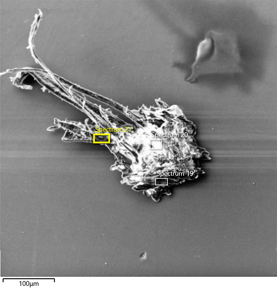
SEM (Scanning Electron Microscope) image
from
Pfizer vial, August batch.
(Courtesy of Dr. Daniel Nagase)
The Pfizer vials he got for the March analysis were kept at room
temperature for around 2 months, and the Moderna vials for about 1
month, he says.
The vials for the August analysis were kept at room temperature for
about two weeks.
Elements,
Temperature, and Time
Dr. Lisa Morici,
told Health.com in December 2020
that the,
"mRNA rapidly
degrades. There are also enzymes in the environment and all
around us that break down mRNA," explaining why the vaccines
need to be frozen so deeply.
Dr. Michael Palmer,
a microbiologist, told us that since the vaccine samples were,
"left to rot for
about 2 months in a liquid state... we don't know whether any of
the strange shapes that were observed in those samples were
actually present in the native material."
"A lot can happen in that time, and we don't know whether any of
the strange shapes that were observed in those samples were
actually present in the native material," Palmer said.
However, he acknowledged
that the complete lack of phosphorus and of nitrogen in the X-ray
spectra is "potentially important."
"Both the mRNA and
the lipid nanoparticles should contain both of these elements;
and whatever chemical degradation might have happened to the
chemical compounds contained during those two months, the
chemical elements those compounds had consisted of would have
been preserved," Palmer continued.
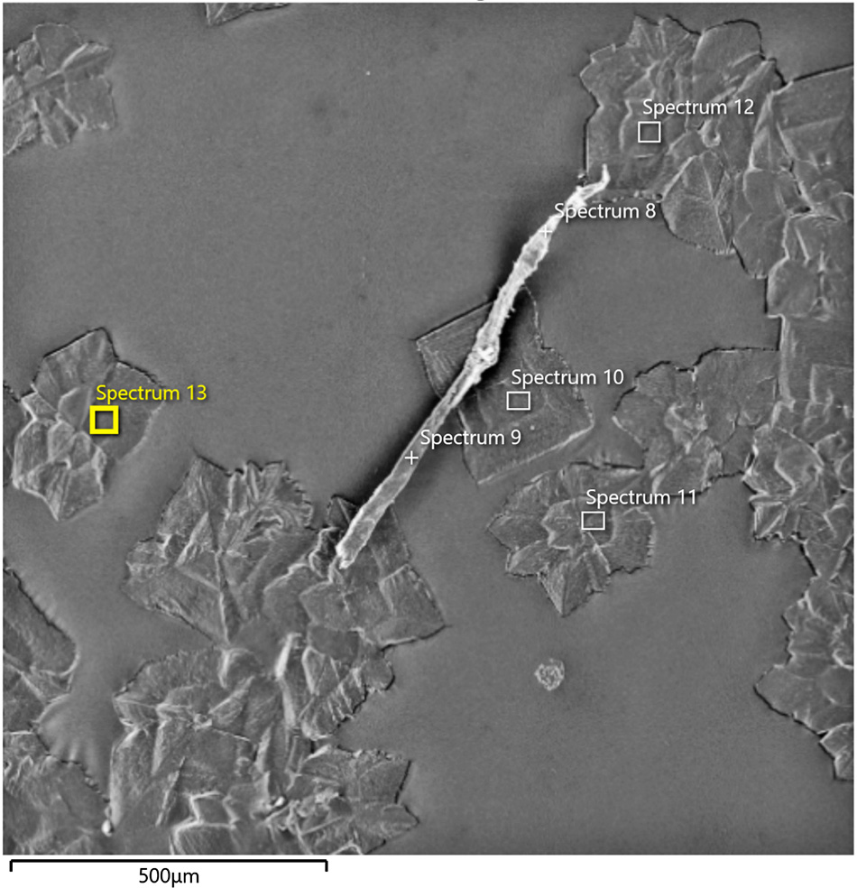
SEM (Scanning Electron Microscope) image
from
Pfizer vial, March batch
(Courtesy of Dr. Daniel Nagase)
"We would need to know, however, exactly what steps were taken
in the preparation of those samples for electron microscopy.
Could it be that the
lipids and the mRNA, or their degradation products, were washed
off, and only the strange shapes remained?
Also, all the spectra
were acquired from the strange shapes, and none from the
background between them.
Thus, based on the
limited information contained in the video, I don't consider the
absence of mRNA and of lipid nanoparticles to have been proven
conclusively," Palmer concluded.
Another microbiologist,
Dr. Sucharit Bhakdi (retired), "partly but not wholly" agreed
with Palmer's point of view.
"The fact is the
'monopoly' of carbon and oxygen in all analyses," Bhakdi pointed
out.
"This ties in with the news of 'empty' batches," he said.
The "bad batch" or "empty
batch" idea refers to the allegations that different batches of
vaccine vials
contain different substances.
SEM (Scanning Electron Microscope) image
from
Moderna vial, March batch
(Courtesy of Dr.Daniel Nagase)
Palmer and Bhakdi recently
published a study about spike
protein expression detection even after 9 months, as well as
inflammation of different organs due to auto-immune response.
"While Dr. Nagase did
not have fresh samples, what he found is consistent with what
everyone else found when they did have fresh samples.
So while we can't say
what those things are, we know they show at a minimum crap
production process," said Sasha Latypova, a former pharma
executive who was on Palmer and Bhakdi's email exchange thread.
About the temperature
issue, Nagase stated his idea:
"The point of
examining unrefrigerated vials is to see what the vaccine does
in conditions more in line with the human body."
He thinks that,
"the refrigeration
temperature does not make any sense."
"Negative 40 [degrees celsius] is not a biological temperature.
Storage of DNA/RNA for several months only requires -20. The
expiry dates for the mRNA vaccines are all 6 months or less
(from what I've seen estimating [based on] the date of
manufacture)," Nagase said.
Scientific
Debate
A recently retired Yale electron microscopist/researcher who talked
to us dismissed Nagase's findings, saying that since the vaccines
were stored in suboptimal conditions, there was "no protocol" for
the research.
"There are no valid
scientific protocols to analyze the contents of vaccines using
these methods.
Furthermore, the
preparation of samples for electron microscopy unfortunately
lends itself to the introduction of artifacts (damage to the
integrity of the sample caused by poor preparation techniques)
which are often misinterpreted under very high magnification,"
the scientist said under the condition of anonymity.
Nagase responded with
this rebuttal:
"The whole point of a
PhD is to write a research paper to prove that you have the
analytical thought process to approach and answer an unknown.
So a proper PhD
committee would evaluate a student's ability to create a
protocol to answer a question where there is no established
protocol.
The critical factor
in determining whether a student has a 'Doctorate' is the
ability for synthesis of knowledge. Creating knowledge where
there was none before."
SEM (Scanning Electron Microscope) image
from
Pfizer vial, March batch.
(Courtesy of Dr. Daniel Nagase)
The former electron microscopist further questioned Nagase's work:
"For example, Dr.
Nagase sees something which seems like a microchip - if you blow
something up hundreds of thousands of times it's hard to
interpret what you're looking at.
It's kind of
analogous to looking at an ink blot because many people have
different interpretations. Some people see a bird or some people
see a tree."
To which Nagase
responded:
"The chip photos are
1,000 to 2,000 x magnification. It says so on the photo. A
scanning electron microscope can't do 'hundreds of thousands.'
That's transmission
electron microscope territory, and if he actually has any
experience with electron microscopy he'd be able to tell from
the nature of the image whether or not it was a scanning or
transmission electron microscope."
The scientist also
questioned Nagase's chain of custody, saying that the images are not
from the vaccines since they have no biological contents.
"I have the chain of
custody, from the provincial health department source of the
samples," Nagase answered.
Other Doctors
Dr. Sherri Tenpeny is a doctor who has been warning about
vaccine dangers for about 30 years.
"Dr. Nagase is one
more in a series of investigators who have discovered unusual
contents inside of a vaccine vial this is supposed to be
manufactured under sterile conditions," Tenpenny told us.
"Researchers
in
Spain,
UK,
Germany,
Japan, and
Brazil who
have viewed multiple
vaccine solutions under a microscope of various magnifications
have all found particulate matter that should not be in any
shot," Tenpenny said.
SEM (Scanning Electron Microscope) image
from
Moderna vial, March batch.
(Courtesy of Dr. Daniel Nagase)
Tenpenny said that there are many articles that refute their
findings.
"Which of course
means they are right over the target," she said.
"No matter what its identification, it should not be injected
into the body and particulates such as this are quite possibly
leading to many of the side effects attributed to the
injections."
'Sine Qua Non
for a Biological Origin'
Dr. James Thorp finds these inclusions in the vials to be
"bizarre" and cannot be "definitively defined."
"I do find it very
peculiar that electron microscopy documents in all of the areas
examined that none of the contents appear to be of biological
origin.
There were no
elements of nitrogen or phosphorus as sampled by electron
microscopic interrogation. Both nitrogen & phosphorus are a sine
qua non for a biological origin," Thorp told us.
"I would not expect the vaccination sample remaining at room
temperature for multiple weeks to test negative for these two
elements that would define a biological source.
Of course, DNA or RNA
would decompose but as a chemistry major, I would not expect the
complete elimination of nitrogen and phosphorus from these old
samples," he said.
"Dr. Nagase's work should not be taken in isolation.
We should consider
that others have found similar 'contaminating' items within the
vials and this should prompt immediate action to investigate
what the 'contamination' is," Dr. Janci Lindsay, a
toxicologist and molecular biologist, told us.
"In the meantime, the inoculation program should be halted. This
would be the logical response if the governments of the world
were truly interested in the health and safety of their
citizens.
The failure to do
that speaks volumes as to the true intent of this program," she
said.
SEM (Scanning Electron Microscope) image
from
Moderna vial, March batch.
(Courtesy of Dr. Daniel Nagase)
SEM (Scanning Electron Microscope) image
from
Moderna vial, March batch.
(Courtesy of Dr. Daniel Nagase)
Pfizer and Moderna did
not respond to a request for comment...
|